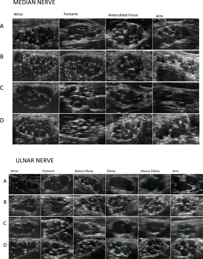
Figure 1.
Median and Ulnar nerve in CIDP (A), in anti-MAG neuropathy (B), d-CIDP (C) and in healthy subject (D). In CIDP (A) thinning of perifascicular epineurium due to compression/dislocation from swollen fascicles. Reduced number of fascicles and swollen fascicles compared to healthy controls especially in more proximal nerve sites due to endoneuronal oedema. Focal/diffuse hypoechogenicity. In Anti-MAG neuropathy (B), reduced number of swollen fascicles with normal or slightly reduced perifascicular epineurium. Same or slightly reduced number of fascicles compared to healthy controls. Normal or slightly reduced echogenicity. In d-CIDP (C), normal number of swollen fascicles with normal or slightly reduced perifascicular epineurium. Normal or focal/diffuse hypoechogenicity.

