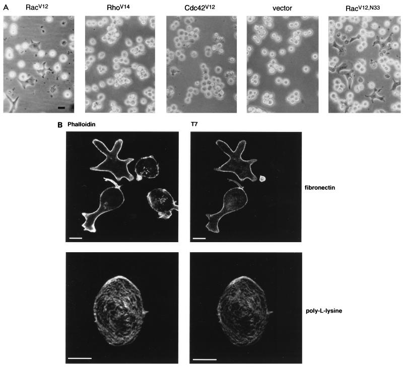
FIG. 1.
Rac induces T-lymphocyte spreading. (A) The Jurkat T-cell line JJK CD4+ was transfected with 5 μg of vector DNA or expression plasmids encoding either RacV12, RhoV14, Cdc42V12, or RacV12,N33 and seeded on fibronectin-coated tissue culture dishes. Six hours posttransfection, cells were visualized using a light microscope and photographed with a Canon camera. (B) RacV12,N33-transfected cells were seeded on fibronectin (upper panel)- or poly-l-lysine (lower panel)-coated coverslips. Cells were fixed and labeled with anti-T7 MAb followed by goat anti-mouse IgG coupled to FITC (right panel). Cells were also stained with rhodamine phalloidin to visualize actin organization (left panel). Only RacV12,N33-transfected cells exhibited the spread phenotype when plated on fibronectin-coated coverslips. Bar = 10 μm.

