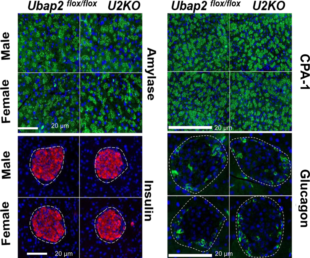
Figure 4. Representative images of pancreatic tissue sections in Ubap2flox/flox and U2KO animals stained for exocrine [Top: (L) Amylase and (R) Cpa1] and endocrine [Bottom: (L) Insulin and (R) Glucagon] genes.

There were no histological differences in the exocrine or endocrine staining between genotypes in mice when stratified by both sex and age. Scale bars: 20μm. Amylase, Cpa1, and glucagon are represented in green. Insulin represented in red. Nuclei were stained with DAPI (blue).
