Fig. 5.
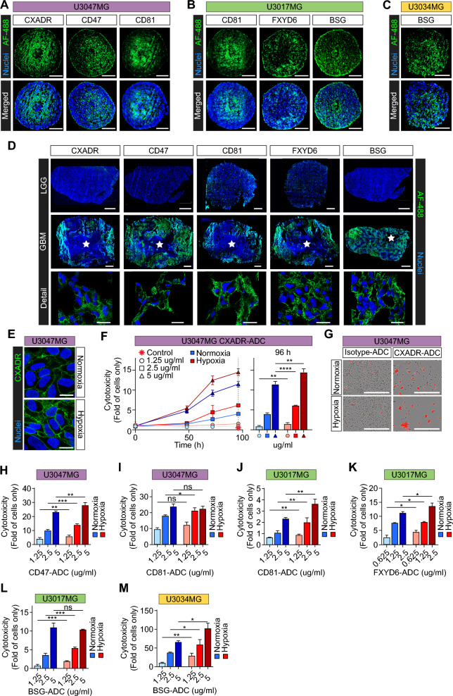
Validation of hypoxia-induced tumor antigen targets for ADC toxicity studies. A–C Representative IF stainings for selected, hypoxia-induced targets, as indicated, in GBM spheroids derived from (A) U3047MG, B U3017MG, and C U3034MG primary cells (scale bars, 200 μm). D Validation of hypoxia-induced targets in GBM and LGG tumors, as indicated (white star: necrosis, scale bars, 1000 and 20 μm for scanned sections and confocal microscopy detail images, respectively). Data shown is representative of at least three patients each. E Increased CXADR protein expression in hypoxic vs. normoxic U3047MG 2D cultures, assessed by confocal microscopy (scale bars, 20 μm). F Left: Cytotoxicity over time by different concentrations of anti-CXADR ADC (isotype control ADC, red star). Cytotoxicity was calculated as red area normalized to confluency, and presented as fold of cells only ± S.D. from 2 independent experiments, each performed in triplicates. Right: Quantification of cytotoxicity at 96 h of treatment. G Representative images from data shown in (F) (5 µg/ml, at 96 h; scale bars, 450 µm). H–M Quantification of cytotoxicity of anti-ADC treatments for 96 h at hypoxia and normoxia, directed against (H) CD47 and (I) CD81 in U3047MG cells; (J) CD81, K FXYD6, and L BSG in U3017MG cells; and M BSG in U3034MG cells. *, **, *** P < 0.05, 0.01, and 0.001, respectively; ns, not significant. IF images are representative of at least 2 independent experiments

