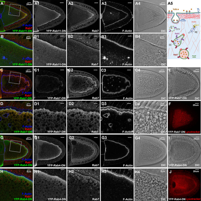
Fig 4. Rab4, Rab5, Rab7 and Rab11 in yolk granule biogenesis.
Confocal fluorescent microscopy and DIC imaging of stage 10 egg chambers with oocyte-specific expression of dominant negative (A-B4) YFP-Rab11-DN, (C-F) YFP-Rab7-DN and (G-J) YFP-Rab4-DN, respectively, that were co-stained by anti-GFP antibody (green), anti-Rab7 (red) and phalloidin for F-Actin (blue), presented as overlaying images in color or as individual channels in gray, or (E, F, I, J) by lysotracker stain alone (red), as annotated. (B-B4, D-D4, H-H4) High-magnification views of the cortex regions highlighted in (A, C, G), respectively, as indicated. Genotypes: The samples were from adult females flies heterozygous for both matalpha4-GAL-VP16 driver (BDSC #7062) and the following UAS-transgenic lines: (A, B) Rab11-DN: UASp-YFP.Rab11.S25N (BDSC #9792). (C-F). Rab7-DN: UASp-YFP.Rab7.T22N (BDSC #9778). (G-J). Rab4-DN: UASp-YFP.Rab4.S22N (#9768). The sizes of the scale bars as annotated inside images.

