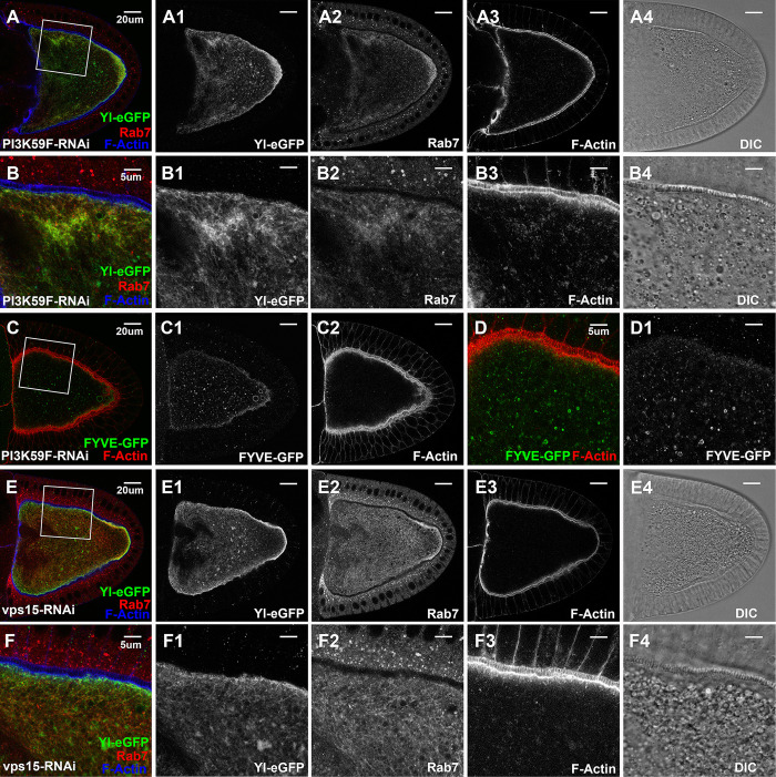
Fig 5. Essential roles of VPS34/VPS15 PI3 kinase complex in Yl recycling and yolk granule biogenesis.
Confocal fluorescent microscopy and DIC imaging of stage 10 egg chambers with oocyte-specific expression of dsRNA again (A-D1) VPS34/PI3K59F or (E, F) Vps15, from flies (A-B4, E-F4) carrying genome-tagging Yl-eGFP-3xHA, co-labeled with antibodies against GFP (green), endogenous Rab7 (red) and phalloidin for F-Actin (blue), or (C-D1) expressing 2xFYVE-GFP, co-labeled for F-Actin (red), presented as overlaying images in color or as individual channels in gray, as annotated. (B, D, F) High-magnification view of the cortex regions highlighted in (A, C, E), respectively, as indicated. Genotypes: The samples were from adult females flies heterozygous for both matalpha4-GAL-VP16 driver (BDSC #7062) and the following UAS-transgenic RNAi and Yl- or FYVE-reporter lines: (A,B) P{TRiP.HMJ30324}attP40 (#64011); p{mini-W+, yl-eGFP-3xHA}. (C,D) P{w(+mC) = UAS-GFP-myc-2xFYVE}2 (#42172); P{TRiP.HMS00261}attP2 (#33384); (E,F). P{TRiP.GL00085}attP2 (#35209)/ p{mini-W+, yl-eGFP-3xHA}. The sizes of the scales as annotated inside images.

