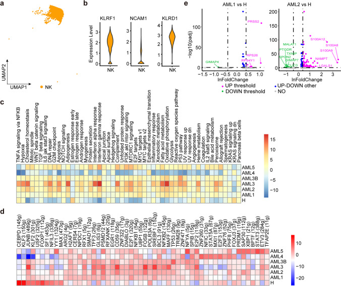
Fig. 4.
Immune landscape of NK cells in AML and healthy samples. a One major NK subcluster was identified by UMAP analysis. b Violin plots show the expression levels of three marker genes in NK cells. c The heatmap of 50 hallmark gene sets in the MSigDB database of NK cells. d Heatmap of AUCell tvalue of transcription factor in NK cells among AML patients and healthy donors. Red and blue represent upregulated and downregulated TFs, respectively. e Volcano plot shows genes differentially expressed in NK cells. Genes displaying significant differential expression are represented by green (downregulated) and purple (upregulated) dots, and selected genes are highlighted

