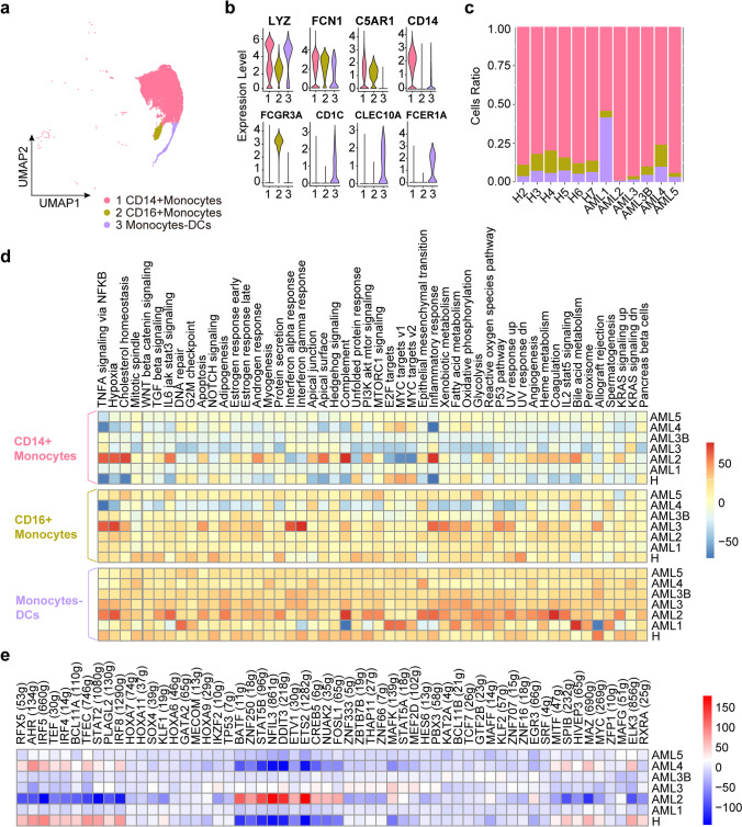
Fig. 5.
Immune landscape of Monocyte and DC cells in AML and healthy samples. a UMAP analysis of CD14+ and CD16+ monocytes identified in the monocytes. b Violin plots show the expression levels of marker genes across the three monocyte subclusters. c Relative proportion of CD14+ and CD16+ monocytes across the monocytes. d The heatmap of the 50 hallmark gene sets in the MSigDB database among the healthy and AML patients. e Heatmap of AUCell tvalue of transcription factor in CD14+ monocytes among AML patients and healthy donors. Red and blue represent upregulated and downregulated TFs, respectively

