Figure 2.
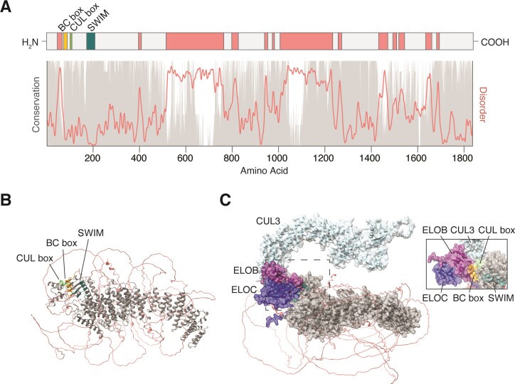
ZSWIM8 protein structure and conservation. (A) Linear diagram of the ZSWIM8 protein showing the size and location of the BC box (orange), CUL box (green), and SWIM domain (teal), as well as several regions of predicted disorder (coral). Below, conservation scores (gray) derived from CONSURF-DB (105) and disorder scores (coral) derived from IUPRED (104) have been plotted along the length of ZSWIM8. In general, intrinsically disordered regions are poorly conserved. (B) The AlphaFold prediction (AF-A7E2V4-F1) of human ZSWIM8 protein is shown (107,108). The BC box (orange), CUL box (green), and SWIM domain (teal) are tightly clustered at one end of the protein and accessible to solvent and other proteins. The conserved protein core (gray) consists of repeating alpha helices that form a solenoid-like structure. The disordered regions (coral) are not confidently predicted. (C) AlphaFold-Multimer prediction (via COSMIC) for ZSWIM8 in complex with ELOB (purple), ELOC (navy), and CUL3 (light blue)(109). The inset image provides another look at the ELOB–CUL3–ZSWIM8 interface from a different angle.

