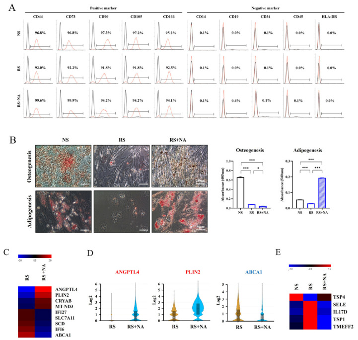
Fig. 4.
Effects of nervonic acid (NA) on stemness, differentiation, and secretion in replicative senescent mesenchymal stem cells (RS-MSCs). (A) Expression of cell surface markers determined using flow cytometry analysis. The x-axis depicts fluorescence intensity, and the y-axis depicts cell count. (B) Osteogenesis was confirmed using Alizarin Red S staining (scale bar=250 μm), and adipogenesis was identified using Oil Red O staining (scale bar=50 μm) in non-senescent mesenchymal stem cells (NS-MSCs), RS-MSCs, and NA-treated RS-MSCs. (C) Heat map shows nine differentially expressed genes (fold change>1.5). (D) Violin plot was used to visualize the difference in expression levels and cell number of three genes in RS-MSCs and NA-treated RS-MSCs (fold change>2). (E) The differences in secretory proteins were confirmed using an antibody array. Heat map shows five differentially secreted proteins (fold change>1.5). Data are presented as mean±SEM. The significance of the differences was assessed by one-way ANOVA (*p<0.05, ***p<0.001).

