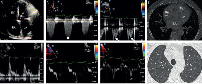
FIGURE 1.
Case 1. Pulmonary hypertension associated with heart failure with preserved ejection fraction: echocardiography and computed tomography (CT). a) Transthoracic echocardiography shows right atrial (RA) and right ventricular (RV) dilatation. In addition, the left atrium (LA) is dilated. Left ventricular (LV) dimensions and systolic function are preserved. b) Maximum tricuspid regurgitation velocity measured by continuous-wave Doppler over the tricuspid valve indicates elevated pulmonary pressures. c) Pulsed-wave (PW) Doppler over the RV outflow tract demonstrates a short acceleration time and a mid-systolic notch (arrows). d) LV diastolic function is severely impaired as illustrated by an increased E:A ratio measured by PW Doppler over the mitral valve inflow. Tissue doppler imaging demonstrates reduced e′ velocity over the e) septal wall and f) lateral LV wall. g) CT of the thorax shows a dilated LA. h) CT shows no signs of parenchymal lung disease.

