FIGURE 2.
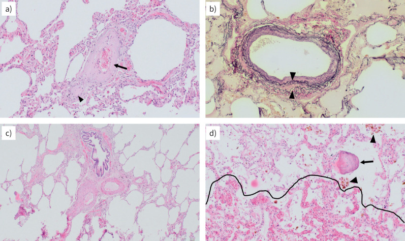
Case 1. Pulmonary hypertension (PH) associated with heart failure with preserved ejection fraction: lung histopathology. a, c, d) Haematoxylin and eosin staining and b) Elastica van Gieson staining; magnification ×100 (all). a) Collagen-rich remodelling with constrictive intimal fibrosis of a septal vein; the small branch (arrowhead) beneath the larger vein (arrow) appears to be completely occluded. b) Arterialisation of a septal vein with duplication of the elastic lamina (arrowheads): these veins resemble arteries, but lack the adjacent bronchioles; note the beige oedema filling the alveoli in part. c) Two pulmonary arterial branches (and their adjacent bronchioles), with substantial intimal and medial thickening and representing the morphological correlate to the pre-capillary PH component. d) Capillary congestion with the beginning of haemangiomatosis-like changes (area below the dotted line); note the numerous haemosiderophages (arrowheads) and the ossified fragment (arrow), both typical in long-lasting congestion due to left heart failure.

