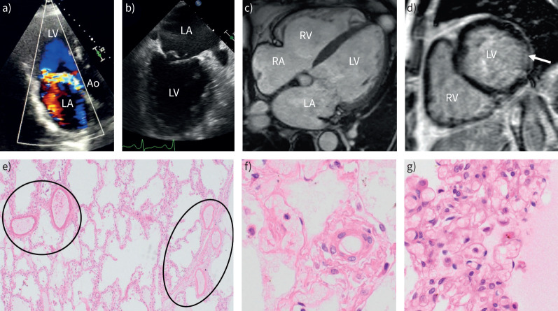
FIGURE 4.
Case 2. Pulmonary hypertension (PH) related to primary mitral valve regurgitation: echocardiography, cardiac magnetic resonance imaging (MRI) and lung histopathology. a) Transthoracic echocardiography illustrates severe mitral regurgitation. b) Transoesophageal echocardiography illustrates a prolapse of the mitral valve. c) Cardiac MRI shows bi-atrial and biventricular dilatation. d) Cardiac MRI with late-gadolinium enhancement demonstrates fibrosis of the left ventricular (LV) inferolateral wall (arrow). e) Lungs from the patient with PH due to mitral valve disease (autopsy). Haematoxylin and eosin staining (all); magnification: e) ×40 and f) and g) ×400. e) Pulmonary arteries (circle on the left) and pulmonary septal veins (circle on the right) are almost indistinguishable due to venous arterialisation; note the thickened alveolar septa in between that are magnified in g). f) Microvessel, either arteriole or venule, of about 30 µm in diameter; note the perfectly round, single-layered muscularisation of this vessel that is under normal conditions totally devoid of smooth muscle cells. g) Alveolar septa are thickened due to congestion and clear capillary haemangiomatosis, similar to capillary changes in pulmonary veno-occlusive disease; note multiple layers of capillaries in a single septum and expanded alveolus to the left and eosinophil fluid within the alveolus on the right side of the alveolar wall (oedema). Ao: aorta; LA: left atrium; RA: right atrium; RV: right ventricle.

