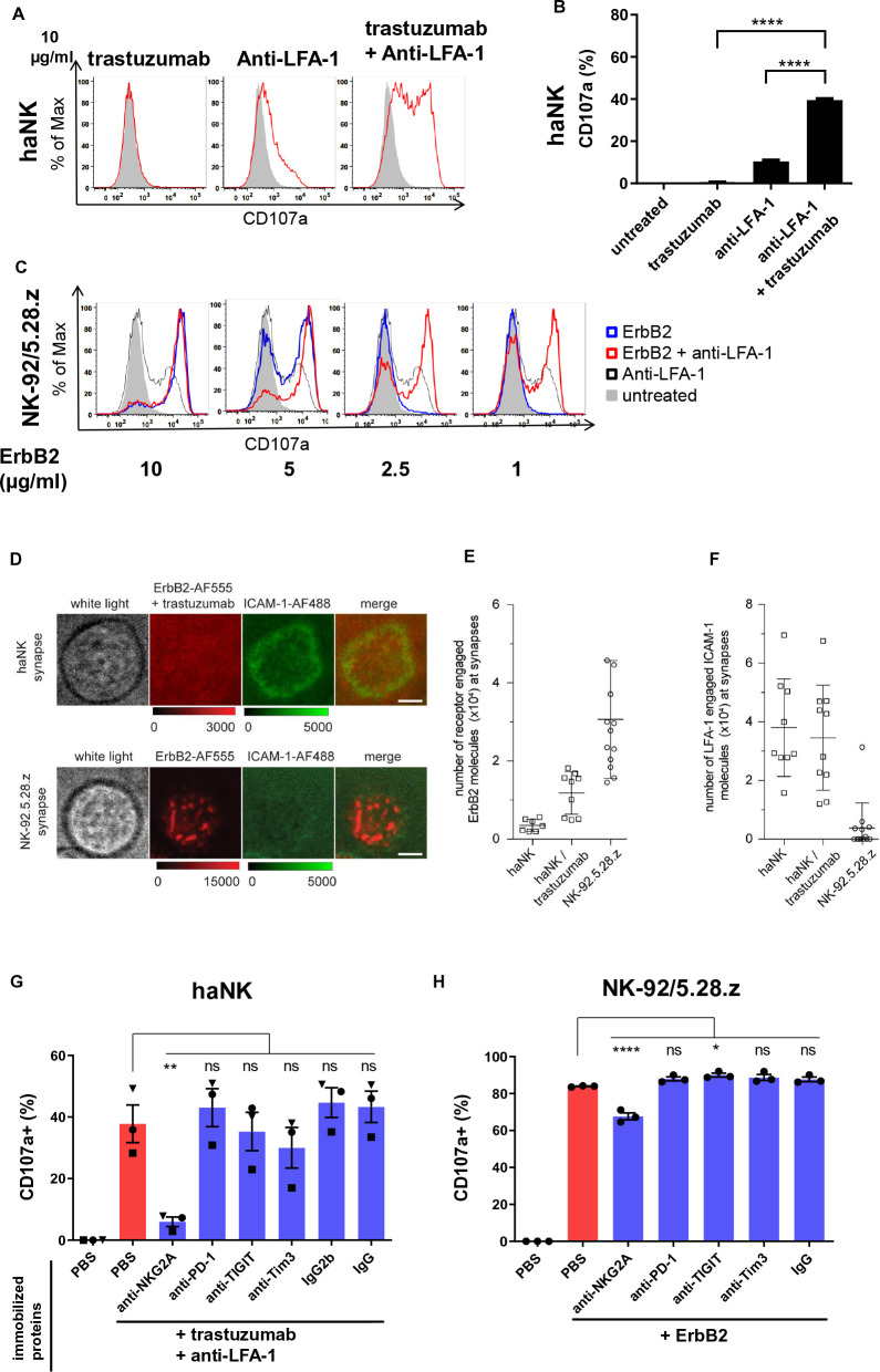
Figure 6.
Chimeric antigen receptor-NK cells form an irregular immunological synapse, which is LFA-1 independent. (A) haNK cells were incubated with indicated plate-bound antibodies in the presence of labeled anti-CD107a antibody for 2 hours, followed by flow cytometric analysis of CD107a as a degranulation marker. Untreated NK cells are depicted in gray. (B) Quantification of CD107a expression. (C) NK-92/5.28.z cells were incubated with plate-bound ErbB2 protein in the presence of labeled anti-CD107a antibody for 2 hours, followed by flow cytometric analysis of CD107a. Representative data from three independent experiments are shown. (D) Total internal reflection fluorescence (TIRF) microscopy images displaying synaptic recruitment of fluorescently labeled ErbB2-AF555 and ICAM-1-AF488 molecules at the synapses of haNK cells in the presence of trastuzumab and NK-92.5.28.z cells. Scale bars: 5 µm. (E, F) Quantification of ErbB2-AF555 (E) and ICAM-1-AF488 (F) molecules recruited at the synapses of haNK and NK-92.5.28.z cells. Each data point represents the number of molecules recruited at the synapse of one cell. Mean values are indicated. Error bars represent±SD. (G, H) haNK cells were incubated with plate-bound trastuzumab and anti-LFA-1 (G) and NK-92/5.28.z with plate-bound ErbB2 protein (H) in each case combined with plate-bound antibodies targeting the indicated inhibitory receptors or immune checkpoint molecules, or respective isotype controls. Degranulation was measured by flow cytometric analysis of CD107a expression. Data were pooled from three independent experiments. Mean values±SEM are shown. haNK, high-affinity FcγRIIIa-modified NK-92 cells; NK, natural killer; PBS, phosphate-buffered saline; PD-1, programmed cell death protein 1; TIGIT, T cell immunoreceptor with Ig and ITIM domains; TIM-3, T-cell immunoglobulin and mucin-domain containing-3.

