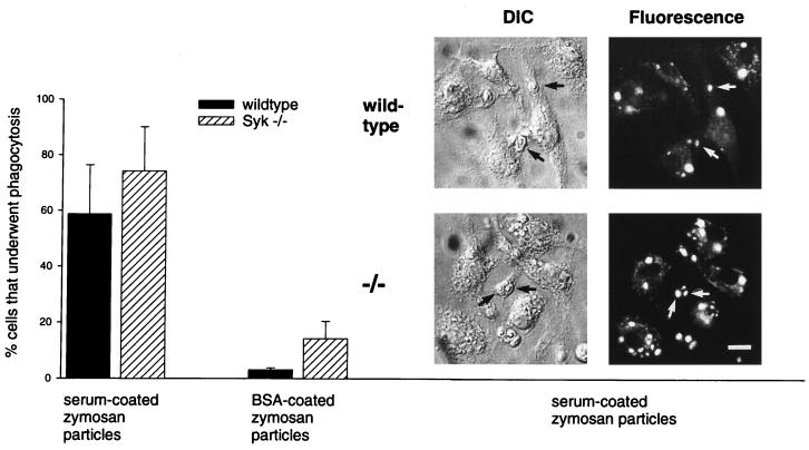
FIG. 8.
Syk−/− macrophages display normal phagocytosis of serum-coated particles. Wild-type and Syk−/− macrophages on glass coverslips were tested for the capacity to phagocytose opsonized zymosan particles in the presence of the fluorescent dye Lucifer Yellow. After 10 min of incubation, successful completion of the phagocytic process was determined by colocalization of zymosan and trapped Lucifer Yellow in sealed phagosomes. The percentage of phagocytosis-positive macrophages is depicted in the bar graph. The photographs show differential interference contrast (DIC) and corresponding fluorescence pictures. Arrows indicate phagocytic events. Bar, 500 μm.

