Fig. 3.
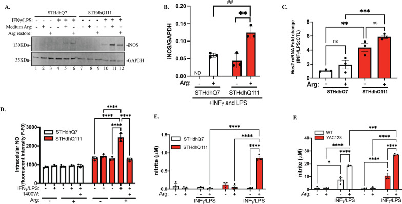
Extracellular Arg regulates IFNγ/LPS-mediated iNOS induction and NO production in HD cells. Cells were first incubated in Arg-free or complete medium overnight. The next day, the cells were treated with IFNγ/LPS for 24 h. To restore Arg under Arg-free conditions, 0.699 mM Arg hydrochloride was added during IFNγ/LPS treatment. A Western blot analysis of iNOS expression in STHdhQ7 and Q111 cells under various conditions as indicated. GAPDH was used as a loading control. B Quantification of iNOS expression in the presence or absence of extracellular Arg during IFNγ/LPS treatment from three independent Western blots by densitometry analysis. **p = 0.0047, ##p = 0.0071, ND not determined. Unpaired t test. C Quantification of changes in Nos2 mRNA expression in the presence or absence of extracellular Arg during IFNγ/LPS treatment by qRT-PCR analysis. **p = 0.031, ***p = 0.0009. D Quantification of intracellular NO levels 24 h after IFNγ/LPS treatment under different conditions as indicated in STHdhQ7 and Q111 cells. ****p < 0.0001. E Quantification of medium NO levels 24 h after IFNγ/LPS treatment under various conditions as indicated in STHdhQ7 and Q111 cells. ****p < 0.0001. F Quantification of medium NO levels 24 h after IFNγ/LPS treatment under various conditions as indicated in WT and HD mouse primary astrocytes. *p = 0.0151, ***p = 0.0007, ****p ≤ 0.0001. In C–F, two-way ANOVA followed by Tukey’s multiple comparisons test. ns not significant

