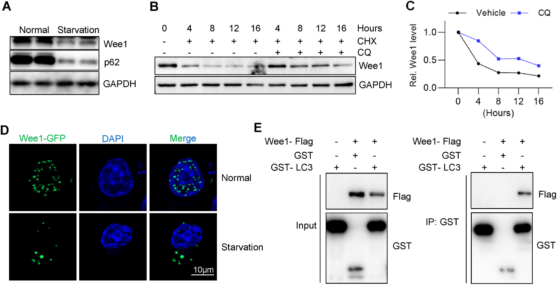
Fig. 3. Wee1 interacts with autophagic receptor LC3 and its turnover is regulated by autophagy.

(A) Cultured HepG2 cells were serum starved for 2 h and subjected to immunoblotting assay. (B) Cycloheximide (CHX) chase assay was performed in the absence or presence of autophagy inhibitor chloroquine (CQ). Briefly, 300 μM of CHX and 200 μM of CQ were used, and the cells were harvested for western blotting at the indicated time. GAPDH was used as the loading control for the immunoblotting assay. (C) Quantification of (B). (D) HepG2 cells stably expressing Wee1-GFP were either treated with normal or serum-free medium for 2 h and then fixed for DAPI staining, followed by confocal imaging. (E) GST co-immunoprecipitation assay demonstrated the interaction between Wee1 and the autophagic receptor LC3. Wee1-Flag, GST, and GST-LC3 plasmids were transfected into HEK 293T cells as indicated, and cells were harvested for IP assay after 36 h.
