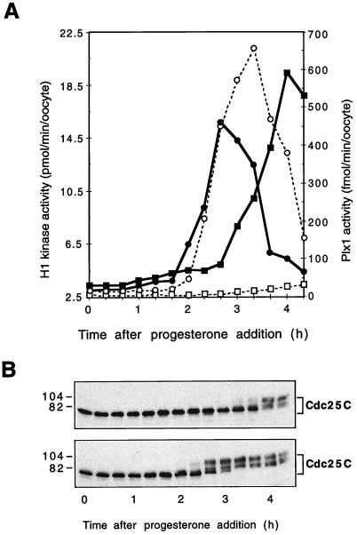
FIG. 4.
Reversal of the inhibitory effect of Plx1 antibodies by Plx1. All oocytes were microinjected with anti-Plx1 IgG (75 ng per oocyte) and then, after 2 h, with either recombinant Plx1 (60 ng per oocyte) or buffer. They were then incubated in the presence of progesterone (3.2 μM), and groups of six oocytes were frozen at the indicated times. (A) Extracts were prepared, and histone H1 kinase and Plx1 activities were determined. Symbols for histone H1 kinase activity: ▪, buffer injection; •, Plx1 injection; Symbols for Plx1 activity: □, buffer injection; ○, Plx1 injection. (B) Samples of extracts were immunoblotted for Cdc25C. Top, buffer injection; bottom, Plx1 injection. Molecular masses (in kilodaltons) are indicated on the left.

