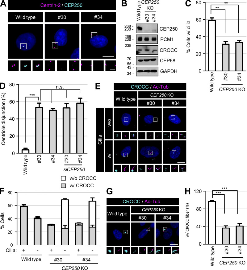
Figure S2.
Generation of the CEP250 KO RPE1 cells. (A) The CEP250 KO RPE1 cells were coimmunostained with antibodies specific to centrin-2 (magenta) and CEP250 (cyan). (B) The CEP250 KO cells were subjected to immunoblot analyses with antibodies specific to CEP250, PCM1, CROCC, CEP68, and GAPDH. (C) The number of cells with cilia was counted. (D) The number of cells with centriole disjunction (>2 μm) was counted. (E) The CEP250 KO cells with and without cilia were coimmunostained with antibodies specific to CROCC (cyan) and acetylated tubulin (magenta). (F) The number of cells with centrosome/basal body CROCC signals was counted in CEP250 KO cells with and without cilia. (G) The CEP250 KO cells were cultured in serum-deprived medium for 48 h to induce cilia assembly and subjected to coimmunostaining analysis with antibodies specific to CROCC (cyan) and acetylated tubulin (magenta). (H) The number of cells with CROCC fibers was counted in cells. (A, E, and G) Scale bars, 10 μm; inset scale bars, 2 μm. (C, D, F, and H) More than 30 cells per group were counted in three independent experiments. Graph values are expressed as mean and SEM. Statistical significance was determined using one-way ANOVA with Tukey’s post hoc test (**, P < 0.01; ***, P < 0.001; n.s., not significant). Source data are available for this figure: SourceData FS2.

