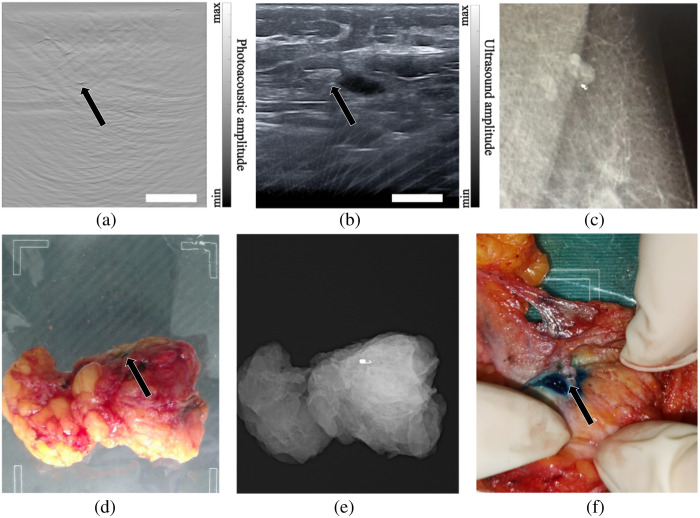
Fig. 6.
Validation of PA/US-based marker detection. (a) PA image of patient 3’s lymph node region; (b) US image of patient 3’s lymph node region; black arrows point to the MC. (c) Mammography shows that the marker was nearby a lymph node. (d) An ex-vivo tumor of patient 4, the black arrow indicates the location of the MB stain. (e) Ex-vivo mammography of the tumor. (f) Photograph of tissue dissection, the black arrow denotes the marker. The scale bar is 10 mm. (See Video 1, MP4, 2.19 MB [URL: https://doi.org/10.1117/1.JBO.29.S1.S11525.s1].)

