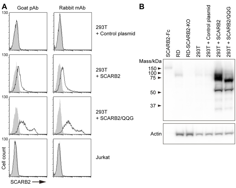
Fig 2. Jurkat cells do not express SCARB2 on the cell surface.
(A) Flow cytometric analysis of cell-surface SCARB2 using goat pAb and rabbit mAb (clone 12H5L1). To confirm both Abs are applicable to flow cytometry, 293T cells overexpressing SCARB2 or mutant SCARB2 with the three amino acid substitutions to enhance cell-surface expression (SCARB2/QQG) and Jurkat cells were stained in parallel. The solid line and the shaded area represent staining with anti-SCARB2 Ab and control IgG, respectively, followed by Alexa Fluor 488-tagged secondary Ab. The figure is representative of three independent experiments. (B) A large amount of overexpressed SCARB2 existed inside the 293T cells. Western blotting analysis with anti-SCARB2 mAb (clone 12H5L1). Recombinant SCARB2-Fc (5 ng) was loaded as a positive control. RD and RD-SCARB2-KO (clone No.3) cells were loaded as positive and negative controls, respectively. The figure is representative of three independent experiments.

