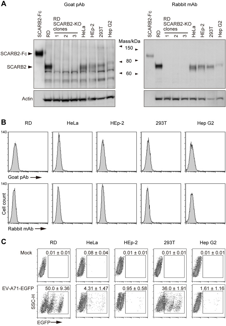
Fig 5. SCARB2 is absent from the cell surface, irrespective of EV-A71 susceptibility.
RD, HeLa, HEp-2, 293T, and Hep G2 were obtained from the ATCC specifically for this study and used after limited passage. (A) Western blotting analysis by anti-SCARB2 pAb (left) and mAb (right, clone 12H5L1). Recombinant SCARB2-Fc (1 ng for pAb, 5 ng for mAb) was loaded as a positive control. RD-SCARB2-KO clones were loaded as negative controls. The figure is representative of three independent experiments. (B) Flow cytometric analysis by anti-SCARB2 pAb (top panels) and mAb (bottom panels), followed by Alexa Fluor 488-tagged secondary Ab. The solid line and the shaded area represent staining with anti-SCARB2 Ab and control Ab, respectively. Note that the solid line and the border of the shaded area are almost completely overlapped, indicating the absence of SCARB2 on the cell surface. Representative results with the following passage numbers after receiving from the ATCC; for pAb: RD, 3; HeLa, 3; HEp-2, 3; 293T, 3; Hep G2, 5; for mAb: RD, 13; HeLa, 5; HEp-2, 4; 293T, 4; Hep G2, 8. As a positive control of SCARB2 staining, cells expressing surface SCARB2 were always stained and analyzed in parallel. The figure is representative of three independent experiments. (C) EGFP expression in cells infected with EV-A71-EGFP. Cells were infected with EV-A71-EGFP at 10 CCID50 per cell and cultured for 18 h. Then EGFP expression was measured by flow cytometry. The EGFP-negative cells are not infected. The majority of EGFP-dim cells were infected early in the incubation period, and are dying and losing EGFP expression; some may have just been infected and are starting to express EGFP. The EGFP-bright cells were infected late in the incubation period are actively producing EGFP. The number indicates the percentage of EGFP-positive cells (mean and s.e. for three independent experiments).

