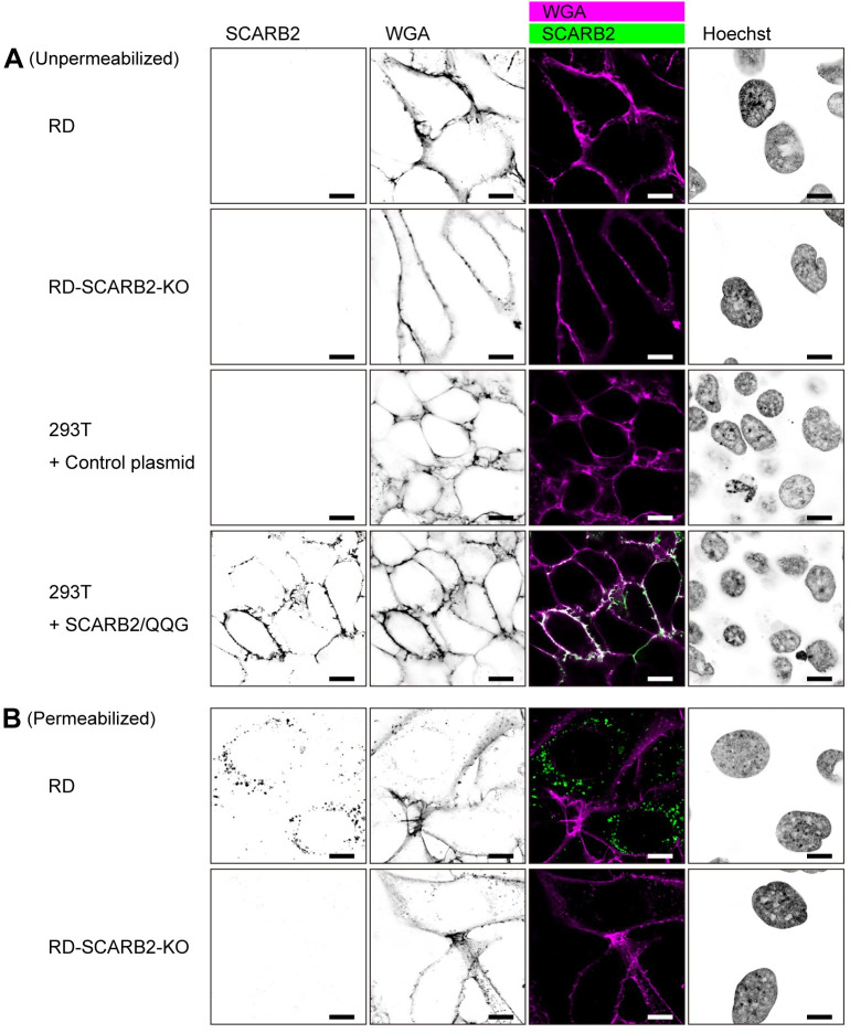
Fig 6. SCARB2 is absent from the cell surface and localized in the cytoplasm of RD cells.
(A) Cells were stained with anti-SCARB2 mAb (clone 22H6L14) followed by Alexa Fluor 488-tagged secondary Ab and WGA conjugated with Alexa Fluor 633 without permeabilization. WGA was used to visualize the plasma membrane. Then the cells were fixed and observed under a confocal microscope. RD-SCARB2-KO cells (clone No.3) and 293T cells transfected with a control plasmid were used as negative controls. 293T cells expressing SCARB2/QQG on the cell surface were used as positive control. The figure is representative of three independent experiments. (B) RD and RD-SCARB2-KO (clone No.3) cells were stained with WGA, fixed, permeabilized, and stained with anti-SCARB2 mAb (clone 22H6L14) followed by Alexa Fluor 488-tagged secondary Ab. The figure is representative of three independent experiments. Scale bars, 10 μm.

