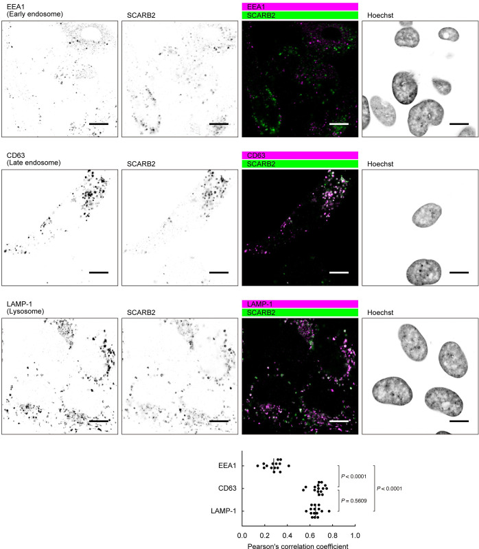
Fig 7. SCARB2 co-localizes with the markers of late endosomes and lysosomes in RD cells.
RD cells were fixed, permeabilized, and stained with anti-SCARB2 mAb (clone 22H6L14) and mAb against either EEA1 (early endosome), CD63 (late endosome), or LAMP-1 (lysosome), followed by Alexa Fluor-tagged secondary Ab. Then the cells were observed under a confocal microscope. The figure is representative of three independent experiments. In each experiment, five pairs of images were analyzed. The graph shows colocalization between the markers and SCARB2 expressed as Pearson’s correlation coefficient. The vertical line indicates the mean value. Scale bars, 10 μm.

