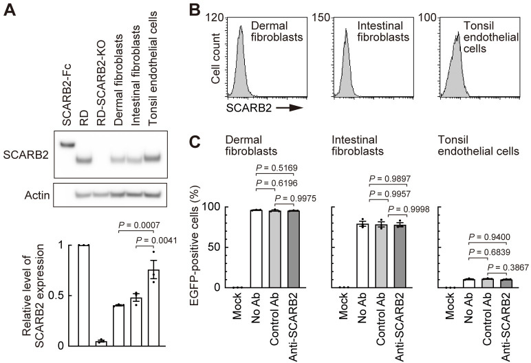
Fig 9. EV-A71 enters human primary cells in a SCARB2-independent manner.
Human dermal fibroblasts (neonatal), intestinal fibroblasts, and tonsil endothelial cells were examined as cells presumed to be involved in the in vivo pathogenesis of EV-A71 infection. (A) Western blotting analysis with anti-SCARB2 mAb (clone 12H5L1). Recombinant SCARB2-Fc (5 ng) was loaded as a positive control. RD and RD-SCARB2-KO (clone No.3) cells were loaded as positive and negative controls, respectively. The figure is representative of three independent experiments. The graph displays the relative level of SCARB2 expression normalized by actin. The relative amount of SCARB2 in RD cells was expressed as 1. (B) Flow cytometric analysis by anti-SCARB2 pAb, followed by Alexa Fluor 488-tagged secondary Ab. The solid line and the shaded area represent staining with anti-SCARB2 pAb and control Ab, respectively. Note that the solid line and the border of the shaded area are almost completely overlapped, indicating the absence of SCARB2 on the cell surface. Representative results of cells passaged twice after receiving from the company. As a positive control of SCARB2 staining, cells expressing surface SCARB2 were always stained and analyzed in parallel. The figure is representative of three independent experiments. (C) EGFP expression in cells infected with EV-A71-EGFP in the presence of anti-SCARB2 pAb at 18 h post-infection. Cells with the following passage numbers after receiving from the company were used; dermal fibroblasts, 4; intestinal fibroblasts, 4; tonsil endothelial cells, 2. Results are indicated as the mean and s.e. for three independent experiments (A, C).

