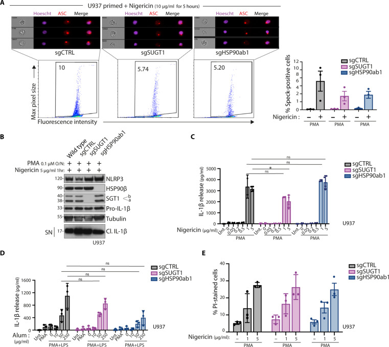
Fig. 4. Stimuli-mediated NLRP3 activation does not rely on the HSP90β-SGT1 pathway.
(A and B) Inflammasome activation in U937 control cells (sgCTRL) or lacking SGT1 (sgSUGT1) or HSP90β (sgHSP90ab1). Cells are treated either with PMA, z-vad-fmk overnight, or nigericin as indicated for 5 hours and intracellularly stained using anti-ASC antibodies and Hoechst to stain the nucleus. Representative images with ASC specks in cells are shown. (A) Cells are analyzed using imaging flow cytometry. (B) Protein expression and release of inflammasome activation markers are measured by Western blot. (C) Inflammasome activation in U937 control cells (sgCTRL) or lacking SGT1 (sgSUGT1) or HSP90β (sgHSP90ab1). Cells are treated with PMA overnight and subsequently stimulated with nigericin for 1 hour. (C) and (D) Inflammasome activation in U937 control cells (sgCTRL) or lacking SGT1 (sgSUGT1) or HSP90β (sgHSP90ab1). Cells are treated with PMA or PMA and LPS, as indicated, overnight and subsequently stimulated with indicated concentrations of nigericin for 1 hour (C) or alum for 6 hours (D). Release of cleaved IL-1β in the supernatant is quantified by ELISA. (E) Inflammasome activation in U937 control cells (sgCTRL) or lacking SGT1 (sgSUGT1) or HSP90β (sgHSP90ab1). Cells are treated with PMA overnight and subsequently stimulated with indicated concentrations of nigericin for 1 hour. Cell death was analyzed by PI-positive staining and measured by flow cytometry. Data are obtained from three independent experiments, represented as means ± SD, and tested for statistical significance using nonparametric Mann-Whitney U test (P ≤ 0.05 is considered significant and indicated with *).

