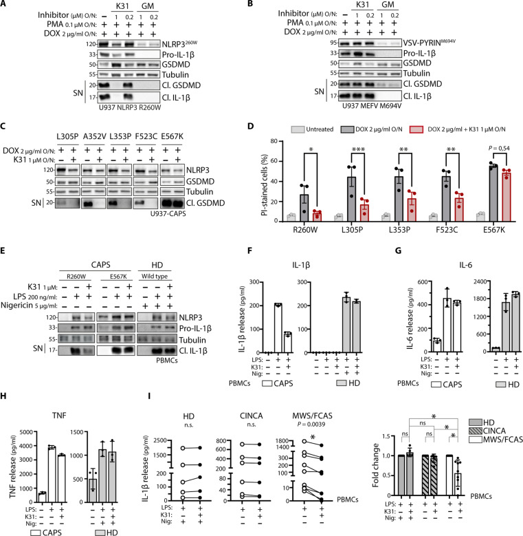
Fig. 5. Pharmacological inhibition of HSP90β reduces IL-1β secretion in CAPS.
(A and B) Cells are treated with PMA and doxycycline overnight in the absence or presence of KUNB31 (K31) or GM. Protein expression and release of inflammasome activation markers are measured by Western blot. Inflammasome activation in U937 cells expressing doxycycline-inducible NLRP3 R260W (A) or MEFV M694V (B). (C and D) Inflammasome activation in U937 cells expressing doxycycline-inducible NLRP3 mutants. Cells are treated with doxycycline overnight. Protein expression and release of inflammasome activation markers are measured by Western blot (C). Cell death in representative CAPS models was analyzed by PI-positive staining and measured by flow cytometry (D). (E to H) Inflammasome activation in healthy donor (HD)– or patient-derived PBMCs. Cells are treated with KUNB31 for 30 min or left untreated, after which LPS is added for 2 hours. For healthy donor PBMCs, nigericin is added for the last 45 min. Protein expression and release of inflammasome activation markers are measured by Western blot (E). Release of cleaved IL-1β (F), IL-6 (G), and TNF (H) in the supernatant is quantified by ELISA. (I) Inflammasome activation in PBMCs derived from healthy donors, CINCA patients, or MWS/FCAS patients. Cells are treated with KUNB31 for 30 min or left untreated, after which LPS is added for 2 hours. For healthy donor PBMCs, nigericin is added for the last 45 min. Release of cleaved IL-1β in the supernatant is quantified by ELISA, shown as raw data or fold decrease. ELISA data are obtained from three independent experiments, represented as means ± SD, and tested for statistical significance using paired t test or two-way analysis of variance (ANOVA) (P ≤ 0.05 is considered significant and indicated with *).

