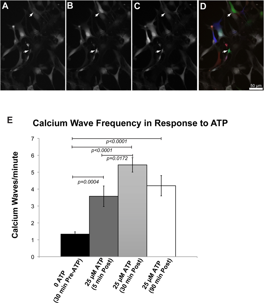
Figure 2. 2-hour imaging of hypothalamic astrocyte cultures using the 3D printed imaging chamber.
Representative images of calcium imaging in hypothalamic astrocytes at the beginning (A), peak (B), and end (C), of a calcium wave in two cells (arrows). The images in panels in A-C, were falsely colored red (A), green (B) and blue (C) and overlaid to make panel D. This panel demonstrates the dynamic nature of signaling, with colored cells representing actively signaling cells, and white cells representing cells between calcium waves. Initial spontaneous background calcium waves in astrocytes represented at 0 ATP (17 cells, 3 biological replicates). After 30 minutes imaging, 25 μM ATP stimulation was preformed via the exchange of fluid in the chamber (19 cells, 3 biological replicates). Imaging was also performed 30 minutes (15 cells, 3 biological replicates) and 90 minutes (10 cells, 2 biological replicates) after this one-time application of 25 μM ATP.

