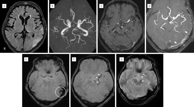
Figure 1.
MRI findings on admission. (A) FLAIR showing edema in the temporal to parietal lobes. (B) MRA MIP 3D reconstruction did not reveal any abnormal vessels. (C) MRA source image. (D) Slab MIP (10 mm thickness) of MRA demonstrating abnormal signal propagating into the TS from the MMA and OA (white arrows). T2*-weighted imaging showed a low signal in the left sigmoid sinus suggesting thrombus (E) (dot circle) and dilatation of the left BVR (F) (white arrowheads), which improved on later images after treatment (G) (white arrowheads). BVR: basal vein of Rosenthal, FLAIR: fluid-attenuated inversion recovery, MIP: maximum intensity projection, MMA: middle meningeal artery, MRA: magnetic resonance angiography, OA: occipital artery, TS: transverse sinus

