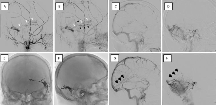
Figure 2.
Cerebral angiography images before and after treatment. Angiography performed before (A-D) and after (E-H) treatment. (A, B) Lateral view of the left external carotid angiography in the early (A) and late (B) phases. Left external carotid angiography showing TSS dAVF with an isolated sinus and shunt blood flow backward from the vein of Labbe (A, B: white arrowheads) to the BVR (B: black arrowheads). (C, D) Before treatment, the StS was not visible in the venous phase on right internal carotid angiography (C) or vertebrobasilar angiography (D), indicating impaired perfusion of the deep veins. (E, F) The isolated sinus was adequately embolized by TAE with ethylene vinyl alcohol (EVOH) polymer (Onyx®, Medtronic, Irvine, USA). (G, H) After treatment, the StS blood flow normalized (black arrows). BVR: basal vein of Rosenthal, StS: straight sinus, TAE: transarterial embolization, TSS dAVF: transverse-sigmoid sinus dural arteriovenous fistula

