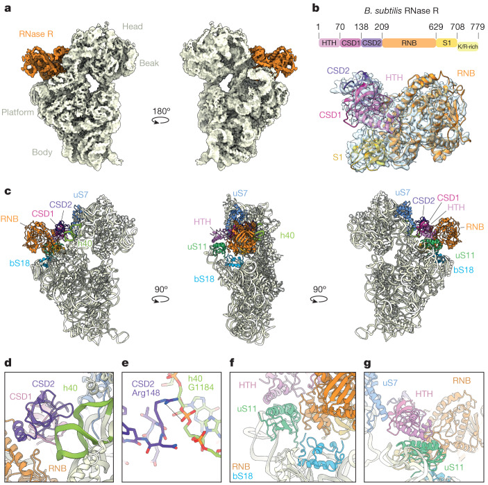
Fig. 1. Interaction of RNase R with the 30S subunit.
a, Two views of the unsegmented cryo-EM map of state I of the RNase R–30S complex, with the density for RNase R (orange) and 30S (pale yellow) coloured. The density has been filtered for visual clarity. b, Cryo-EM map density (transparent) and molecular model for RNase R, with RNase R domains coloured according to the schematic (HTH domain, pink; CSD1, magenta; CSD2, purple; RNase II family 3′ exonuclease domain (RNB), orange; S1 domain, gold; and K/R-rich tail, yellow). c, Overviews of the RNase R-binding site on the 30S subunit, highlighting interactions with 30S components h40 (green), uS7 (blue), bS18 (sky blue) and uS11 (lime). For RNase R, individual domains are indicated and coloured as in b. d, Interaction of the CSD2 domain (purple) with h40 (green). e, Interaction of Arg148 of CSD2 with G1184 in h40. f, Interaction of the HTH domain (pink) and the RNB domain (orange) with uS11 (lime) and bS18 (sky blue). g, Interaction of the HTH domain (pink) and the RNB domain (orange) with uS11 (lime) and uS7 (blue).

