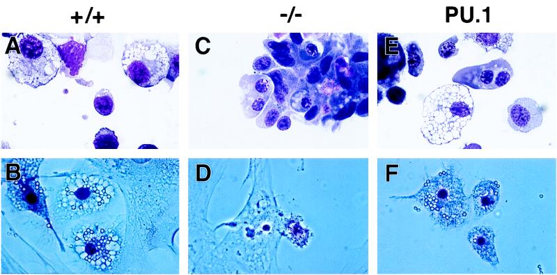
FIG. 3.
Representative cells from day 11 EB cultures. MGG-stained hematopoietic cells in cultures derived from PU.1+/+ ES cells (+/+), PU.1−/− ES cells (−/−), and a PU.1−/− ES cell clone transformed with the wild-type PU.1 transgene (PU.1) are shown. Macrophages capable of phagocytizing latex beads (after 4 h at 37°C) are clearly apparent in the PU.1+/+ and rescued cultures and absent from the PU.1−/− cultures. Light-field photomicrographs are depicted in panels A, C, and E, while phase-contrast photomicrographs are shown in panels B, D, and F. Magnification, ×69.

