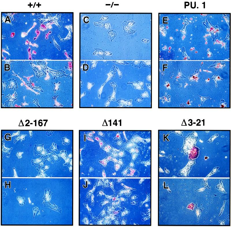
FIG. 4.
Identification of macrophages by immunocytochemical staining. Phase-contrast photomicrographs of staining by antibodies for CD11b (A, C, E, G, I, and K) and F4/80 (B, D, F, H, J, and L) are shown for differentiated PU.1+/+ ES cells, PU.1−/− cells, and transformants containing wild-type (PU.1), Δ2-167, Δ141, and Δ3-21 cDNAs. Magnifications: A to J and L, ×36; K, ×90.

