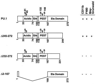
FIG. 6.
Diagram of wild-type and deletion mutant PU.1 cDNAs tested in the ES cell system. Numbers indicate the positions of amino acid residues in PU.1; the acidic, glutamine-rich, PEST, and Ets-type DNA-binding regions are represented as boxes. Serine residues thought to be phosphorylated are also indicated. PURI constructs capable of promoting myeloid development were scored for the presence of CD11b+ or F4/80+ cells or cells with the morphology of macrophages upon MGG staining.

