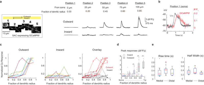
Fig. 4. Direction-selectivity of calcium transients emerges in the distal half of SAC dendrites.
a Left: ROIs (#1–5) of a SAC dendritic branch expressing GCaMP6f. Right: dF/F0 traces in ROIs on the left (Mean and SEM). b Overlay of Vm from current clamp recordings and GCaMP6f traces (Mean, SEM). c Normalized GCaMP6f responses at different distances from the soma along individual dendritic branches. Each line represents data from a single branch. The maximum response of each branch is normalized to 1. An abrupt increase of GCaMP6f signal is detected in distal half of the dendrite in the outward direction. d Binned dF/F0 and SEM of response amplitude at different fractions of dendritic radius. Kolmogorov-Smirnov Test, **p < 0.01, n = 11 cells. e Summary of rise time and half width of fast outward GCaMP6f transients in the distal half of dendrites. Bin size: 0.4–0.6; 0.6–0.8; 0.8–1. n = 11 cells. For each box in (d, e), the central mark indicates the median, and the bottom and top edges of the box indicate the 25th and 75th percentiles, respectively. The whiskers extend to the most extreme data points not considered outliers, and the outliers are plotted individually using the ‘+‘ symbol. Source data are provided as a Source Data file.

