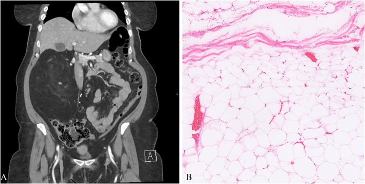
Figure 1.
A, Computed tomography scan shows a large, 14.6 cm fat-containing mass in the right retroperitoneal, pararenal space with slight enhancement of a small solid component. (B) “Giant” lipoma arising in the retroperitoneum showing typical features of mature adipocytes with thin fibrous strands traversing through fat lobules (patient 5).

