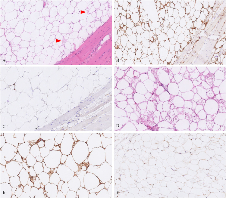
Figure 3.
A, Tumor from patient 3 showed an intramuscular retroperitoneal lipoma-like hibernomas with scattered rare brown fat cells (arrowheads). (B) CD10 showed strong staining in this example. (C) CD68 is shown in the same areas, which helps to discriminate from histiocytes. (D) The tumor from patient 6 showed prominent brown fat cell population with strong CD10 staining (E) as compared to negative or faint positivity in ordinary retroperitoneal lipoma without brown fat (F).

