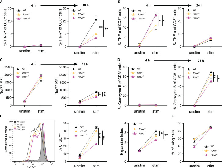
Figure 5.
P2X4 and P2X7 are involved in the expression of cytokines and activation markers as well as the induction of proliferation. (A–D) Spleen cells from WT, P2rx4 -/- and P2rx7 -/- mice were incubated with and without anti-CD3 mAb and anti-CD28 mAb (A, C) or only anti-CD3 mAB (B, D). After 4 h and 18 h or 24h, the expression of IFN-γ (A), TNF-α (B), Nur77 (C) and granzyme B (D) was analyzed by FACS given as % of CD8+ T cells (A, B, D) or mean fluorescence intensity (MFI) of antibody staining (C). (E) Spleen cells from WT, P2rx4 -/- and P2rx7 -/- mice were labeled with CFSE and incubated with and without anti-CD3 mAb and anti-CD28 mAb. Representative results for CFSE staining of CD8+ T cells is shown in the histogram. The strength of proliferation is given as % CFSElow and as expansion index which takes into account the number of divisions. (F) Survival of cells was estimated by % of living cells after culture for 72h with and without stimulation. Mean ± SEM, N = 3 per group. (F) Results were analyzed with two-way ANOVA and Tukey’s post-test (*p < 0.05; **p < 0.01; ***p < 0.001; ****p < 0.0001).

