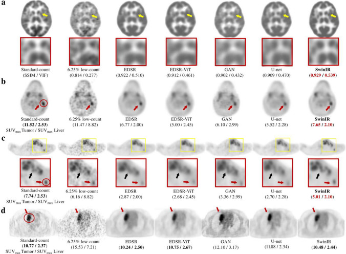
Fig. 6.
Examples of denoised 18F-FDG PET images by emerging approaches. Each column (a) to (d) indicates different patients or organs. From left to right, the sample images show the standard-count PET, low-count PET, and denoised PET images, corresponding to the enhanced deep super-resolution network (EDSR), EDSR-ViT, GAN, U-Net, and Swin Transformer image restoration network (SwinIR).
© 2023 SNCSC. Reprinted with permission from Wang et al. [159]

