Figure 4.
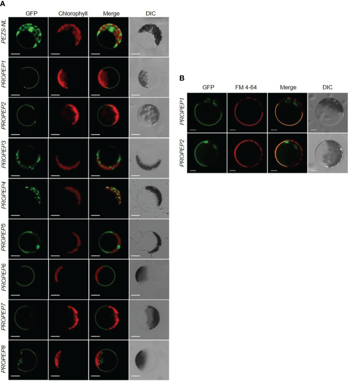
The subcellular localization assay of PROPEPs. (A) Arabidopsis mesophyll protoplasts were transiently transformed with a PEZS-NL vector expressed GFP signaling as the control. The coding sequence without the stop codon of PROPEP1 to PROPEP8 were cloned into pEZS-NL vector and transiently transformed into Arabidopsis mesophyll protoplasts. Columns from left to right show GFP signals (GFP), chlorophyll autofluorescence (Chlorophyll), merged images of GFP and chlorophyll (Merge), and bright-field differential interference contrast (DIC). Bars =5 μm. (B) FM 4-64 signaling was co-expressed with PROPEP1-GFP and PROPEP2-GFP in Arabidopsis protoplasts. The PROPEP1 and PROPEP2 fused GFP protein were transiently transformed into Arabidopsis mesophyll protoplasts and stained with 1 μM FM4-64 for 15 s before photographed. Columns from left to right show GFP signals (GFP), FM 4-64 fluorescence signals (FM 4-64), merged images of GFP and FM 4-64 (Merge), and bright-field differential interference contrast (DIC). Bars =5 μm.

