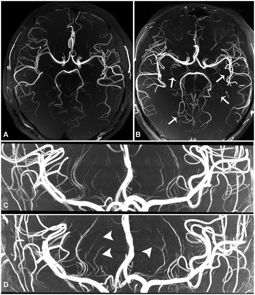
Figure 1.
Normal volunteer scan comparison. 3-Tesla 3D TOF-MRA with 0.44 mm isotropic voxel size, acquisition time of 10 minutes 21 seconds (a), shows fewer lenticulostriate and perforator branches than what is seen on 7-Tesla 3D TOF-MRA with 0.3 mm isotropic resolution, acquisition time of 7 minutes 14 seconds (b, arrows). 7-Tesla 3D TOF-MRA with 0.3 mm isotropic resolution (c) compared to 7T TOF-MRA with 0.2 mm resolution, acquisition time 10 minutes 14 seconds (d). There are an increased number of visualized lenticulostriate branches (arrows) at higher resolution acquisition (d, arrowheads), though with use of compressed sensing there is increased noise artifact due to reduced signal, resulting in small branch irregularity.

