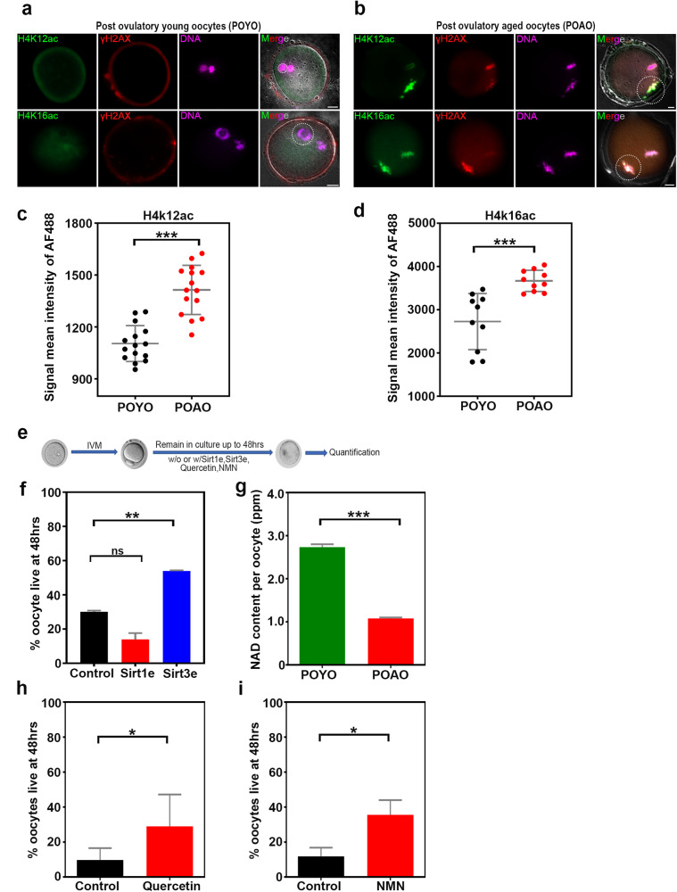
Fig. 5. Epigenetic modifications and NAD metabolism regulates the POAO death.
a, b POYO and POAO stained for histone modifications H4K12 or 16 ac (green), DNA damage marker γH2AX (red) and DNA (magenta). c, d Quantification of H4K12, 16ac signal intensities in POYO and POAO. e Schematic representation of postovulatory oocyte ageing and treatments (f) Quantification of % live POAO in the presence and absence of SIRT1 and SIRT3 enhancers. g Quantification of NAD+ in POYO and POAO. h, i Quantification of % live POAO in the presence and absence of CD38 inhibitor quercetin and NMN. 20–25 oocytes used for H4K12 and 16ac localization. 80 to 100 oocytes used for treatments. Error bars show mean ± s.e.m ***P ≤ 0.0001, **P ≤ 0.004, *P ≤ 0.03, (n.s.) P ≥ 0.01, paired t-tests. Scale bars = 10 μm.

