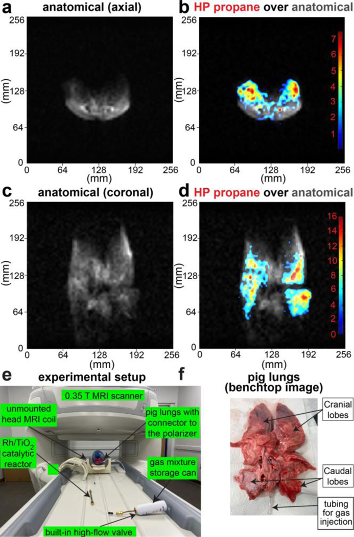
Figure 5.
Fast slice-selective 2D GRE images of HP propane gas injected in the excised pig lungs and recorded with 64×64 imaging matrix over 256×256 mm2 FOV, 50-mm slice thickness, and 30-degree slice-selective RF pulse. a) Axial anatomical image of the inflated excised pig lung, b) Corresponding axial false-color image of HP propane gas contrast agent overlaid over greyscale axial anatomical image shown in display a, c) annotated photo of the experimental setup showing the gas mixture tank, Rh/TiO2 reactor, unmounted head 0.35 T MRI coil and gas connection from the reactor outlet to the lungs’ trachea. d) Coronal anatomical image of the inflated excised pig lungs, e) Corresponding coronal false-color image of HP propane gas contrast agent overlaid over greyscale coronal anatomical image shown in display d. f) Annotated photo of the excised pig lungs employed for the pilot imaging studies. Each image in displays a, b, d, and e was acquired in 0.94 s, see SI for additional images and details.

