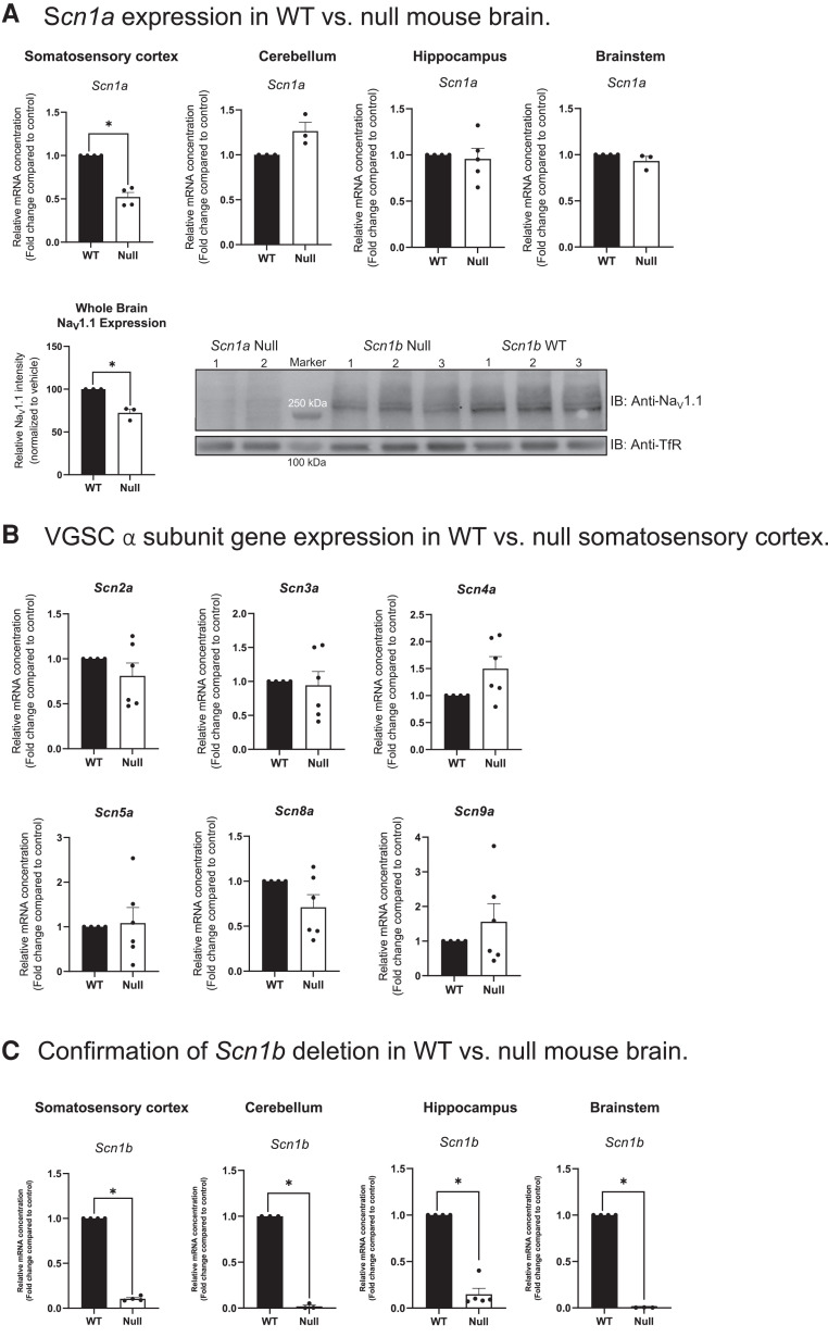
Figure 4.
Differential VGSC α and β subunit expression in P15-18 Scn1b+/+ and Scn1b−/− mouse brains. (A) Scn1a and Nav1.1 expression in Scn1b+/+ (WT) versus Scn1b−/− (null) mouse brain. Scn1a gene expression was significantly decreased in null somatosensory cortex (*P < 0.001); however, no changes in Scn1a were detected in the cerebellum, hippocampus or brainstem (P > 0.05). Bottom panel: Nav1.1 protein expression was significantly decreased in Scn1b null mouse whole brain membranes compared with WT whole brain. Left: Quantification of anti-Nav1.1 immunoreactive bands normalized to corresponding anti-TfR bands for Scn1b WT versus null brains for the blot shown on the right. Data are represented as means ± SEM for three WT and three null brains, respectively. Statistical significance was determined using Student’s t-test (*P < 0.01). Right: Western blot analysis of Nav1.1 protein in Scn1b null and WT whole brain membranes, as indicated. Upper blot: anti-Nav1.1. Lower blot: anti-TfR. Molecular weight markers are indicated. (B) VGSC α subunit gene expression in WT versus null somatosensory cortex. No changes were detected in the relative expression of Scn2a, Scn3a, Scn4a, Scn5a, Scn8a or Scn9a between null and WT somatosensory cortex (P > 0.05). (C) Confirmation of Scn1b deletion in WT versus null mouse brain. Relative expression of Scn1b in null and WT mouse somatosensory cortex, cerebellum, hippocampus and brainstem (P < 0.0001). Statistical significance was determined using Student’s t-test (P-value < 0.05). Data are represented as the mean ± SEM. WT: n = 3–5, null: n = 3–5. Male and female mice were used in all experiments (A and B).

