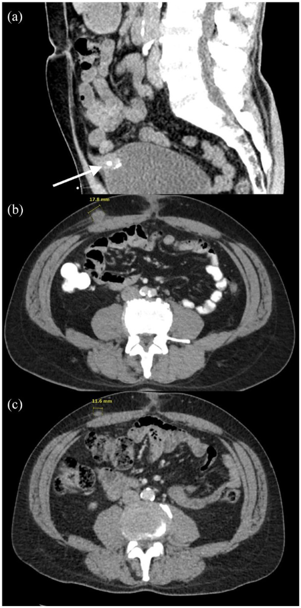
Figure 2.

CT of the abdomen demonstrates urachus. (a) Sagittal view, respectively, of 2.7 × 2.0 cm peripherally calcified urachal remnant with vesicourachal diverticulum stone with respective arrows at the time of initial hematuria and right flank pain. (b) Axial CT imaging denoting peripherally enhancing subcutaneous mass along the right anterior abdominal wall 4 months after partial cystectomy and excision of urachal mass. (c) Axial CT views of the abdomen demonstrating interval decreased size of metastasis of peripherally enhancing subcutaneous mass along the right anterior abdominal wall after one cycle of Gem-FLP.
CT, computed tomography; FLP-Gem, gemcitabine, cisplatin, 5-FU, and leucovorin.
