
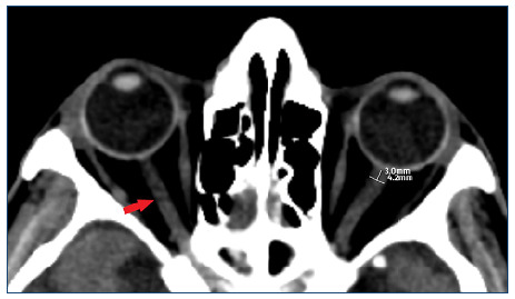
Figure 1. In the axial computed tomography section, the right optic nerves (red arrow) (a) and the location where the left optic nerve sheath diameter measurement was done (b).
Official websites use .gov
A
.gov website belongs to an official
government organization in the United States.
Secure .gov websites use HTTPS
A lock (
) or https:// means you've safely
connected to the .gov website. Share sensitive
information only on official, secure websites.