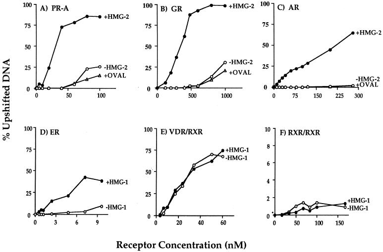
FIG. 5.
Saturation DNA binding analysis of purified nuclear receptors in the presence or absence of HMG-1/-2. The concentrations of purified receptors indicated in the figure were varied in DNA binding reactions against a constant amount (0.3 ng) of 32P-labeled oligonucleotide probe, and DNA binding was analyzed by EMSA. Each receptor was assayed alone (−HMG-1 or −HMG-2) or with the addition of 150 to 300 ng of HMG-1, HMG-2, or ovalbumin (+OVAL). The oligonucleotide probe for PR-A (panel A), GR (panel B), and AR (panel C) was a palindromic GRE-PRE. A palindromic ERE oligonucleotide was used as the probe for ER (panel D), a DR-3 oligonucleotide was used for the VDR-RXR heterodimers (panel E), and a DR-1 probe was used for the RXR homodimers (panel F). Purified receptors were quantitated by protein Bradford assay and by comparing silver-stained receptor band intensities with those of known amounts of purified molecular weight protein standards. The specific DNA complexes at each concentration of receptor were quantitated by phosphorimage analysis and plotted as the percentage of total DNA.

