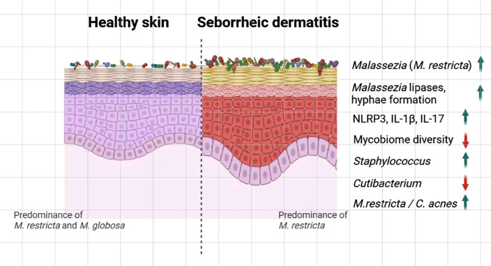
FIG. 2.
Graphical depiction of altered microbiome in skin affected by seborrheic dermatitis (SD). Studies demonstrated reduced diversity of fungal communities and increased abundance of Malassezia restricta in SD-affected skin than in healthy skin. Studies also suggested increased expression of lipases and hyphae formation in Malassezia. Activation of the NLRP3 inflammasome leading to IL-1β secretion and increased IL-17 cytokine production was also observed in the SD skin lesion. Furthermore, bacterial microbiome analysis demonstrated increased abundance of Staphylococcus, while Cutibacterium was reduced. The higher M. restricta/C. acnes ratio in scalps affected by SD compared to that in healthy scalps was also reported. The image was prepared using Biorender.com.

