


Fig. 21.
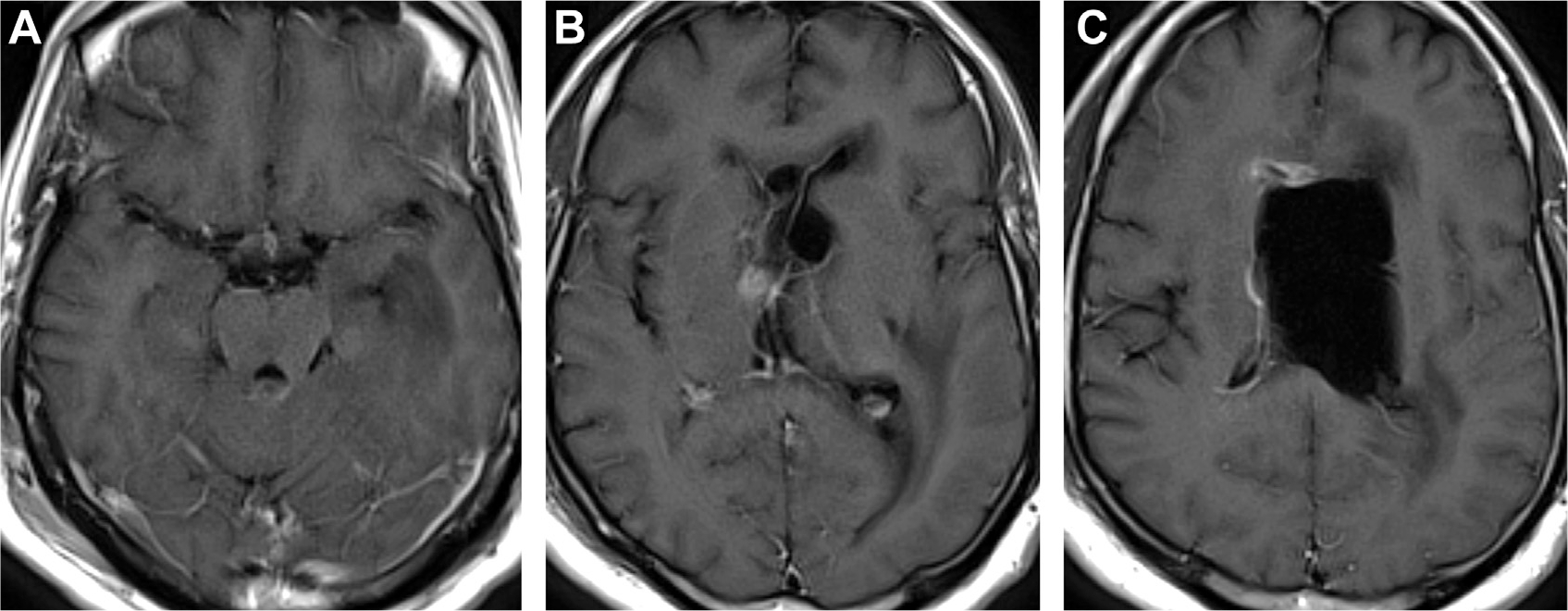
Cryptococcosis and hydrocephalus. T1 postcontrast MR imaging visualizes subtle perimesencephalic and bilateral posterior leptomeningeal enhancement (A). Enhancement along an obstructed foramen of Monro (B). Ependymal enhancement with enlarged left lateral ventricle (C). The right lateral ventricle is reduced in volume due to left ventricle compression and drainage by a ventriculostomy catheter.