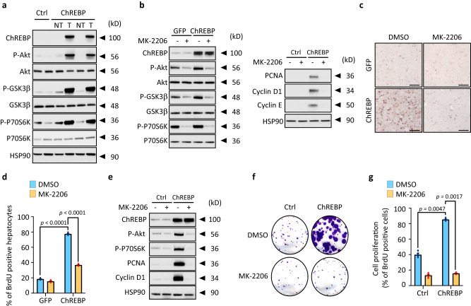
Fig. 3. ChREBP overactivation stimulates the pro-oncogenic PI3K/AKT signaling.
a Representative Western blot analysis of the activity of the PI3K/AKT signaling in ChREBP tumors (n = 10 biologically independent mice per group). b, c Mice, injected with either GFP or ChREBP overexpressing adenovirus, were orally treated with MK-2206. Captisol (30%) was used as a vehicle for the drug and the control animals were treated with vehicle only. MK-2206 (120 mg/kg) was given orally for 3 weeks on alternate days. b Western blot analysis of the PI3K/AKT signaling and proteins of the cell cycle (n = 6 biologically independent mice per group). c Representative staining of liver sections with BrdU. Scale bars = 100 μm (d) Quantification of BrdU staining is shown (n = 6 biologically independent mice per group). e–g HepG2 cells, stably overexpressing ChREBP, were treated with MK-2206 (100 nM) for 24 h. e Representative Western blot analysis of proteins of the PI3K/AKT signaling and cell cycle (n = 3 independent experiments). f Representative clonogenic assay shown (n = 3 independent experiments). g Cell proliferation index determined by measuring the % of BrdU positive cells (n = 3 independent experiments). All error bars represent mean ± SEM. Statistical analyses were determined by two-way analysis of variance (ANOVA) and Tukey’s multiple-comparisons test. Source data are provided as a Source Data file.

